


Important Foliar & Viral Diseases in Sugarbeet
Sugarbeets are an important crop in Montana. The sugarbeet root is full of sucrose that can be extracted to produce pure sugar. In order to produce the highest percentages of sugar, sugarbeets must remain healthy.
Last Updated: 12/17by Jessica Rupp, Ph.D., MSU Extension Plant Pathologist; and Barry Jacobsen, PhD, MSU Professor Emeritus
SUGARBEETS ARE AN IMPORTANT CROP IN
Montana, and across many regions of the United States. Sugarbeets are produced in 14 states under both dryland and irrigated conditions. This equates to around 1.5 million acres of sugarbeets grown annually. This produces nearly 4 million tons of sugar. In Montana, 45,000 acres of irrigated sugarbeets are planted annually, which produces almost 1.6 million tons. The growing area in the state follows the Yellowstone river stretching from around Billings, Montana, to the northeast corner of the state. The sugarbeet root is full of sucrose that can be extracted to produce pure sugar. In order to produce the highest percentages of sugar, sugarbeets must remain healthy. Identification and management of sugarbeet diseases is critical to ensure optimal yield.
Cercospora leaf spot
General Information
Cercospora leaf spot (CLS), caused by the fungus Cercospora beticola, is considered the most destructive foliar disease of sugarbeet. It is the most important foliar disease of sugarbeet in Montana. Yield losses can approach 40% or more under conducive environmental conditions. In the field, lower harvest weight is common and sugar percentage is reduced. Beets with lower sugar content don’t store as well as those with higher sugar content. In storage, decay takes place, which can lead to increased molasses content and greater levels of impurities. Losses are attributed to the loss of leaf area to the lesions, and the toxins, cercosporin and beticolin produced by the fungus. As the sugarbeet invests more energy to produce more leaves, sugars are unable to be stored in the root.
Symptoms and Spread
Cercospora leaf spot produces round lesions that are very small, 3-5 mm in diameter. Spot centers appear light brown to brown (Figure 1). Upon close inspection, black fungal stroma may be present within the lesion. In high humidity conditions, whitish conidia and conidiophores may be present on the stroma. As the disease progresses the lesions may coalesce (Figure 2). When leaves die, they remain attached to the plant. The toxins produced by the fungus cause rapid leaf death. Spots can also be found on petioles, where they may appear more elongated. Full leaf death of those leaves supplying most of the energy for sugar production can occur in serious infections (Figure 3).
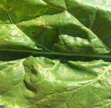
FIGURE 1. Early infection of sugarbeet with CLS. Lesions are small and round with a dark brown border. (Photo by Jessica Rupp)
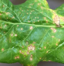
FIGURE 2. Cercospora lesions coalesce if the disease is allowed to progress. (Photo by Jessica Rupp)
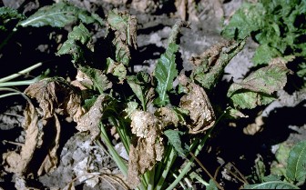
FIGURE 3. Older leaves die due to Cercospora infection, thereby vastly reducing the plant’s ability to store energy in the form of sugars in the root. (Photo by Howard F. Schwartz, Colorado State University, Bugwood.org)
The fungus survives between seasons in infected leaves and petioles as conidia and pseudostroma. Conidia can persist up to four months on infected leaf debris. Pseudostroma can persist up to two years. Other sources of inoculum can include infested seeds and wild hosts. Conidia require water films, or very high humidity and temperatures ranging from 68°F to 79°F. Under favorable conditions, conidia usually spread via rain splash or irrigation. Wind, insects, equipment, or humans can all spread conidia. Conidia are produced from primary infections in 7-21 days. The disease is most damaging in warm, humid summers. A full disease cycle can take place in as few as 10 days.
Management
Crop rotations of at least three years allow for the decay of any debris left in the field. Destroy weeds and any leftover debris prior to planting. Cultivation can speed debris degradation. Fungicides are available to control the disease. Use proper timings and full rates. Alternate fungicides to manage fungicide resistance development to fungicide groups. When possible, use varieties with genetic resistance. Distances of at least 300 feet are recommended between the current and previous sugarbeet fields. Do not plant into fields used the previous season for sugarbeets. Scouting should begin prior to the onset of row closure and continue throughout the season. Levels of susceptibility differ among varieties. If growers have had Cercospora leaf spot issues in previous years, they should consider a more tolerant variety.
Models
Many models exist to help growers predict the environmental conditions for action, as well as yield loss components. Daily potential infection values (DIVs) are reported and based on the number of hours per day where humidity is greater than 90 percent and the recorded hourly temperatures. Using the model criteria, these two values are then expressed as a number between 0-7. Two days values are added together to create a DIV of 0-14. Values of 7 or above indicate high risk for the disease to develop and often indicate that intervention is required. In Montana, values between are considered conducive for disease development. When values reach 4-6, it is critical to scout the crop. When values meet or exceed 7, and the pathogen is present it is time to consider action, even if no spots have been detected on leaves. It is critical to apply a fungicide when conditions first favor disease. Late application of the first spray often leads to difficult season-long control regardless of subsequent fungicide application timings.
Fungicides and Fungicide Resistance
Resistant isolates of Cercospora beticola are present in states surrounding Montana. Currently, no resistant isolates have been found in Montana. Because of this, it is very critical to rotate fungicide mode of action, both in season, and into the following season. When discussing fungicide resistance, it is important to understand that this designation means that the fungus is unaffected by the fungicide that previously gained control in the field. This differs from the term tolerance. In this instance tolerance means that the fungus growth is reduced under a level of fungicide that previously prevented fungal growth. If tolerant strains are found in the field, growers can expect a reduced level of control. If resistant strains are present in the field, growers will see no control. If you suspect either scenario, please contact MSU Extension or Schutter Diagnostic Lab.
It is recommended that growers be especially aware of CLS resistance to the benzimidazole class of fungicides; although not found yet in Montana. Because the potential development of fungicide resistance in this class is particularly high in Montana, MSU recommends that a tank mix be used with a benzimidazole (thiophanate methyl) and TPTH (triphenyltin hydroxide). Mix according to label instructions. Labels may be found at www.cdms.net. Research from North Dakota State University indicates that this fungicide combination works best when used as the first foliar fungicide application. This application is to prevent the disease and is applied prior to identifying CLS in the crop. DIVs are reported by the Sugar Co-op agriculturalist in the region and should be used as the indicator of disease risk. Agricultural staff will provide advice on the timing of the first spray application when environmental conditions are right for the disease. Never apply the same fungicide(s) or fungicide classes consecutively. Use at least ¾ rate of all fungicides applied in a tank mix.
Instances of CLS isolates tolerant to TPTH have been around since the early 1990s in eastern sugarbeet growing areas. This can be very challenging to identify at the field-scale as it can be difficult to tell if the cause is fungicide application problems or tolerant isolates. Lastly, resistance has been reported to the QoI fungicides, the strobilurins. Products containing active ingredients in this family, especially pyraclostrobin should be vigilant about rotation of mode-of-action and field scouting. Resistant isolates were detected in North Dakota in 2016.
QoIs, Benzimidozoles, Triazoles, Ethylenebisdithiocarbamates (EBDC), and TPTH products are registered for use for controlling Cercospora leaf spot. Consult product label for rates and pre-harvest intervals. Tank mixing of fungicides has proven to be a valuable approach to management.
For more information on fungicides, please see CDMS (www.cdms.net), the North Dakota State University fungicide guide or the NDSU sugarbeet production guide (www.ag.ndsu.edu/publications/), the MSU Extension MontGuide Fungicide Use in Field Crops: Classification, Risks, Use & Economics (MT201705AG) and other sources.
Phoma leaf spot
General Information
The disease is caused by a seedborne fungal pathogen, Phoma betae. While the pathogen most often causes root rot symptoms (Figure 4) both in the field and during storage, Phoma leaf spot presents as distinctive spots visible on leaves. The disease is considered of little direct economic importance, it remains important as a source of inoculum for seed production, and for awareness of potentially contaminated seed.
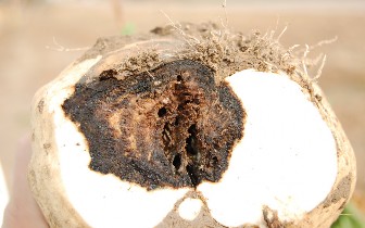
FIGURE 4. Root rot symptoms. (Photo by Robert Harveson, University of Nebraska)
Symptoms and Spread
Phoma leaf spot produces leaf spots that are usually 1-2 cm in diameter. The spots themselves contain patterns of concentric rings, differentiating them from other leaf spot pathogens. Black pycnidia, small in size, develop in the ringed lesion. Phoma betae is a fungus that produces pycnidiospores, also known as conidia, that are black. During the fall, perithecia develop on the underside of leaves. These perithecia are also black. Ascospores, yellow-green in appearance, may also be present.
The fungus is seedborne and can survive as conidia or as mycelium in soil for over two years. In wet conditions, the pycnidia can take on water, then exuding a gelatinous mass of spores, which can then be disseminated by water. Perithecia are commonly dispersed by wind later in the season. This disease is most prevalent under high humidity and warm temperatures. Identification is aided by digging up the root and slicing it open to reveal symptoms.
Management
The best management techniques involve the use of a four-year rotation of sugarbeet with non-host crops. Producers should control the weed lambsquarters. One of the most powerful management options is the use of treated seed.
Alternaria leaf spot
General Information
Alternaria leaf spot is a common disease of sugarbeet. It is generally of little importance in Montana, but continues to persist at low levels. Its presence with other diseases, such as viruses, often leads to confusion and misdiagnosis.
Symptoms and Spread
Lesions on leaf surfaces begin as small pinpricks, and expand in diameter. The lesions may be circular or irregular in shape, ranging from gray to dark brown (Figure 5). These lesions then break, causing holes in the leaf surface termed “shot-holes.” Lesions often begin on older leaves. These two pathogens are often found in fields that already have some infection of the Beet yellows virus complex. When present with viruses, lesions begin at the interveinal region. Two different fungi, Alternaria brassicae and Alternaria alternata cause Alternaria leaf spot. Both of these fungi have a wide host range, including members of the cruciferous vegetable family, members of the Solanaceae family including potato and tomato, and the grass family.
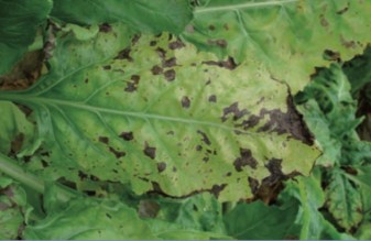
FIGURE 5. Alternaria alternata infecting sugarbeet leaves. (Photo by IBRAB)
The disease cycle of these two fungal pathogens is not well-studied in sugarbeet. It is known that inoculum persists in the debris of infected plants, leading to the assumption that both pathogens can also overwinter on sugarbeet debris. Spores are produced on leaf lesions and are then dispersed by the wind. High humidity often favors sporulation, followed by periods of lower humidity ideal for spore dispersal. Cool, humid conditions favor infection of new hosts. A. alternata often colonizes plant wounds.
Management
Disease management is often unnecessary in sugarbeet due to the usual timing of infection. Infections often occur very late in the season, corresponding with leaf senescence. Often, the disease is well-controlled with irrigation and fertility management and regular fungicide applications.
Powdery Mildew
General Information
Powdery mildew has been a disease of variable importance in sugarbeet since its first identification in the late 1930s. Until the mid-1970s the disease had a low level of importance, but in 1974 there was an outbreak of significance in several western states. Since then, the disease has occurred to some degree in all sugarbeet growing regions.
Symptoms and Spread
The pathogen first appears as a whitish mat, small and often circular in size on lower, older leaves (Figure 6). Given ideal conditions, the mildew can spread rapidly to all leaf surfaces. The leaf tissue underneath can appear yellow to purplish-brown. On a whole field scale, a blue cast may be apparent. Left unchecked, powdery mildew can kill the plant. Additionally, infected leaves are more susceptible to frost damage. Mildew may also spread to the stems. Powdery mildew on sugarbeet is caused by Erysiphae polygoni. Specific morphology of the fungus is influenced by weather conditions. Conidia are produced and arise from mycelia growing on the leaf surface. Ascocarps can form within the lesion and are dark brown to black in appearance, resembling pepper granules.
In moderate climates, the disease can overwinter, but in Montana, inoculum often is carried northward on wind during the spring, summer, and fall. Conidia travel on the wind, landing on new beet crops. The conidia germinate, and mycelia grow on the leaf surface. More conidia are produced, which serve as sources of secondary inoculum. During favorable conditions, the fungus forms ascospores. The disease commonly begins in the south, under favorable conditions that allow the fungus to overwinter. Then, the disease moves northward and eastward, often from California. Optimal temperatures for disease development ranges in the mid-to-upper 70s F. Within-field conidia production and secondary infections are most common under low relative humidity. Production of ascospores increases with increased disease severity. Frequent rains or sprinkler irrigation impedes disease progress. Susceptibility in sugarbeets increases with plant age.
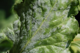
FIGURE 6. Powdery mildew on sugarbeet. (Photo by Oliver T. Neher, The Amalgamated Sugar Company, Bugwood.org)
Management
Management of the disease is recommended if the disease is present more than one month prior to harvest. Control measures should be applied at the first sign of disease. Repeat applications may be required.
Rhizomania: Beet necrotic yellow vein virus
General Information
Beet necrotic yellow vein virus (BNYVV) is the causative agent of Rhizomania, a common viral disease affecting sugarbeets worldwide. No variety has complete resistance to the virus, however, many varieties carry a dominant gene that confers a strong tolerance to the virus. Rhizomania has a fairly narrow host range, including sugarbeets.
Symptoms and Spread
Early infection of sugarbeet in the field results in stunted plants that often display visual symptoms, while older plants sometimes remain asymptomatic. Early infections can reduce both sugar yield and quality. Plants that may be older, exhibiting severe root infections, may not always have foliar symptoms. The occurrence of the symptoms for which the virus is named, necrotic yellow vein is now rare in the field. Therefore, diverse criteria need to be evaluated. Symptoms mid-season can often be mistaken for nitrogen deficiency. Fluorescent yellow foliar symptoms are common. When a Rhizomania-tolerant variety is grown in the field, a symptom commonly called “blinkers” is often apparent. Blinkers are areas of fluorescent yellow foliage, growing in an unusually upright format, characteristic of infected plants (Figure 7). These same plants also exhibit root symptoms, as well (Figure 8). Root symptoms are characterized by light brown discoloration of the root, easily confused with other diseases. Diagnostic tests are necessary. Early infection results in root stunting and “root bearding,” the over-production of secondary and tertiary roots.
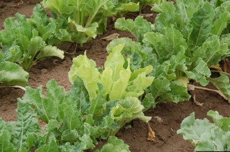
FIGURE 7. Blinkers, caused by Rhizomania. (Photo by Oliver Neher, The Amalgamated Sugar Company, Bugwood.org)
Later infections often result in characteristic “wine- glass” shaped roots and bearding of the lower root. Beet necrotic yellow vein virus is a member of the Benyvirus family. This virus has multiple rod-shaped particles, and most often uses vectors for dissemination. The virus is transmitted by the protozoan, Polymyxa betae. Polymyxa betae is capable of forming spores that can survive long periods in the soil under adverse conditions. When the spore germinates to infect a plant, the virus is transmitted as well and can begin its own replication. The virus can also be mechanically transmitted.
Management
Currently, the best means of disease management is the use of genetic resistance. The ‘Holly’ gene, also called Rz, is a dominant gene with the best available resistance to BNYVV to date. Other management practices include ensuring good soil drainage, limiting irrigation in known areas of concern, early planting, avoidance of soil movement, and harvesting in dry condition.
Beet Curly Top Virus
General Information
Beet curly top virus (BCTV) is now considered the first serious viral disease of sugarbeet. It is widespread throughout the sugarbeet growing regions in the western United States. The virus is also prevalent in Europe, the Middle East, and Asia. BCTV was one of the first diseases determined to be caused by a virus and has a very wide host range.
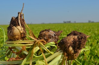
FIGURE 8. Rhizomania, caused by Beet necrotic yellow vein virus, also results in stubby roots. (Photo by Bill Wintermantel, USDA-ARS)
Symptoms and Spread
BCTV causes plants to have a “curly” appearance (Figure 9). Dwarfed, crinkled leaves have a tendency to roll upward and inward, producing a curly pattern. Veins on the underside of the leaf become thickened and on occasion, spiney. Young roots are stunted and often have a distorted, twisted appearance. Rootlet death leads to rootlet regrowth, giving the sugars a hairy-root appearance. The virus also causes phloem tissue in the roots to become necrotic, which then develops cracks. Phloem can be exuded onto the leaves and stems.
BCTV, a single-strand DNA virus, is a member of the curtovirus genus in the family Geminiviridae. There are three main viruses that cause Curly top. In addition to Beet curly top virus, Beet severe curly top virus (BSCTV) and Beet mild curly top virus (BMCTV) occur and are both more common in the U.S. The three viruses often occur as a complex, and can participate in viral recombination with each other, which can lead to greater diversity and increased risk for the development of more aggressive strains. The viruses are all transmitted by Circulifer tenellus, the beet leafhopper (Figure 10). The virus is considered persistent in the vector, meaning the leafhopper can transmit the virus for an extended period, up to one month after only feeding on an infected plant for a short time. The vector, which is native to the western United States, has a wide host range and a high reproductive capacity. It can also migrate over great distances during the growing season. The severity of infection often depends on environmental factors that affect both the crop and vector.
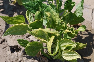
FIGURE 9. Beet curly top virus infecting sugarbeet. (Photo by Howard F. Schwartz, Colorado State University, Bugwood.org)
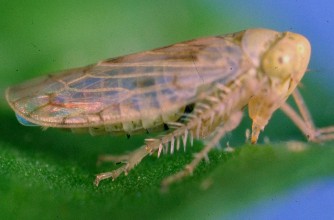
FIGURE 10. Beet leafhopper, the vector of Beet curly top virus. (Photo by A.C. Magyarosy, Bugwood.org)
Management
Insecticides are the most widely used methods to control this virus by management of the vector. These are often applied to sugarbeet as a seed treatment, or an in-furrow treatment. Early planting is favored in some areas to allow growth prior to leafhopper infestations. Controlling weeds along the field edges can also aid in slowing the disease. Tolerant sugarbeet varieties exist.
Glossary
Ascocarp: a fungal fruiting body that contains ascospores.
Ascospores: a spore formed within an ascus.
Ascus: a sac in which spores develop.
Beticolon: a toxin produced by the fungus Cercospora beticola which causes leaf death.
Cercosporin: a toxin produced by the fungus Cercospora beticola which causes leaf death.
Conidia: a spore produced asexually by a fungus.
Conidophores: the fungal structure which bears conidia.
Mode of Action: a biochemical mechanism by which a pesticide has activity against a pest of interest.
Mycelium: the vegetative part of a fungus that is made up of branching filaments called hyphae.
Resistant Fungal: pathogen growth is not prevented by the chemical. This can be due to innate resistance or acquired resistance to the fungicide applied.
Persistent (virus transmission): The virus that can circulate through the entire vector. Vectors acquire the virus in hours to days, and can transmit up to a few weeks. The virus is retained through molting, but is not passed to offspring.
Perithecia: a round or flask-shaped fruiting body of a fungus with a pore through which spores are ejected into the environment.
Petioles: the stalk that joins the leaf to a stem.
Pseudostroma: A false stroma; a cellular body that resembles a stroma.
Pycnidia: an asexual fruiting body produced by a fungus; pycnidium.
Pycnidiospore: a conidia formed inside a pycnidium.
Stroma: a mass of fungal tissue, having spore-bearing structures in or on the surface.
Vector: an organism that transmits a disease from one plant to another by biting, cutting, sucking, or probing.
Viral recombination: the exchange of genetic information between related viruses that leads to new viral strains.
Additional resources
For more information on sugarbeet disease management, see the NDSU Sugarbeet Production Guide, https://www.ag.ndsu.edu/publications/crops/browse-by-crop/. For fungicide label information, please see www.cdms.net/Label-Database.